Here is a sample, how to write a field visit report for BSc. Microbiology 4th year Tribhuvan University Nepal.
Abstract
Introduction
Objectives
List of photographs
Instrumentations
For leishmaniasis
Reference
Our thank and appreciation also go to my friends in developing the report and people who have willingly helped me out with their abilities.
DOTS program is being run in association/collaboration with the National Tuberculosis program and involving Municipality of Dharan and District Health Office, Sunsari.
The main functionally running instrumentations that we have seen in BPKIHS are as follows –
BPKIHS has also been envisioned by the Nepali Parliament as a center of national importance to produce skilled health workforce to meet the country’s need and also to function as a center of excellence in the field of tropical and infectious diseases. Its best example is the ongoing Kala-azar Project in collaboration with international bodies.
In the laboratory, through the Department of Microbiology, various bacteriological, parasitological, mycological, mycobacterial and serological tests are also done along with the CD4 count service. Culture sensitivity, staining procedures, Microscopy, Stool occult blood, Blood for Malaria parasites and microfilaria, Bone marrow/splenic aspirate for LD bodies, Optimal, rk39, DAT, and a wide range of immunological services for different diseases are performed.
For Leishmaniasis
Laboratory diagnosis of leishmaniasis can be made by the following:
The following laboratory investigations and diagnostic tests will be done for diagnosis of Kala-azar and PKDL and be monitoring the progress and side effects of the drugs during the course of treatment.
Rapid dipstick test (rK39 test):
rK39 is a 39-amino acid repeat that is part of a kinesin-related protein in Leishmania chagasi and which is conserved within the Leishmania donovani complex. Among the available serological diagnosis of VL, only the immunochromatographic rK39 assay can be considered a point of care test for field application. They are currently the best available diagnostic tool for VL for use in remote areas, and their use in a field setting should be promoted. Apart from use in routine services, use of rK39 in campaigns and active case search is highly recommended. This test may be positive in a healthy person from the endemic areas (asymptomatic cases, past infection). Such a case should be followed up and treated only when they have indicative signs and symptoms of Kala-azar. rK39 is usually negative amongst PKDL with macular lesions and Leishmaniasis.
rK39 is available at Kala-azar endemic districts from level II and above health institutions.
Diagnosis of PKDL All suspected cases of PKDL should be confirmed with the rK39 test. However, it is important to note that the rK39 test may be negative in PKDL cases with only macular lesions and Kala-azar/HIV coinfection. If there is strong suspicion amongst rK39 negative suspected cases, such patients should be referred to centers where the diagnostic facility of PCR or demonstration of the parasite in pinch biopsy is available.
The PCR-ELISA technique can potentially be used for the diagnosis of VL from peripheral blood samples.
– Chemical methods that inactivate the amplicon’s ability to be templates in a new cycle of PCR.
In BPKIHS PCR lab use a spectrum of these methods to effectively control contamination.
Principle:
It is a chain reaction, a small fragment of the DNA section of interest needs to be identified which serves as the template for producing the primers that initiate the reaction. One DNA molecule is used to produce two copies, then four, then eight and so forth. This continuous doubling is accomplished by specific proteins known as polymerases, enzymes that are able to string together individual DNA building blocks to form long molecular strands. To do their job polymerases require a supply of DNA building blocks, i.e., the nucleotides consisting of the four bases adenine (A), thymine (T), cytosine (C) and guanine (G). They also need a small fragment of DNA, known as the primer, to which they attach the building blocks as well as a longer DNA molecule to serve as a template for constructing the new strand. If these three ingredients are supplied, the enzymes will construct exact copies of the templates.
Applications:
A new latex agglutination test (KATEX) for detecting leishmanial antigen in urine of patients with VL. The antigen is detected quite early during the infection and the results of animal experiments suggest that the amount of detectable antigen tends to decline rapidly following chemotherapy. The test performed better than any of the serological tests when compared to microscopy.
Specific antibodies can also be detected by Western blotting. For this type of testing, promastigotes of L. donovani are grown to log phase and lysed and the soluble protein is run on sodium dodecyl sulfate-polyacrylamide gel electrophoresis gels. The separated proteins were electroblotted onto a nitrocellulose membrane and probed with serum from the patient.
Specimens:
The specimens for diagnosis of tuberculosis are,
Carbol fuchsin acid-fast stain or fluorescent auramine-rhodamine stain.
Principle:
The fluorescence microscope is based on the principle of fluorescence. Fluorescence is a phenomenon in which certain fluorescent chemicals absorb the light of a particular wavelength and then emits the lights of larger wavelength with the lesser content of energy.
The specimen is illuminated with the light of a specific wavelength which is absorbed by the fluorophore, causing them to emit the light of longer wavelength. The excited light is separated from the emitted fluorescence through the use of spectral emission filter. The filters and diachronic mirror are selected to match with spectral excitation and the emitted fluorescence of the fluorophore used to label the specimen. In this way, the fluorescence of a single fluorophore is detected in the form of an image at a time. Multicolored images of several types of fluorophore can be detected by using a combination of the filter.
Application:
The fluorescence microscope is an important diagnostic tool for academic and pharmaceutical research, pathology, and clinical medicine.
The test is a molecular test which detects the DNA in TB bacteria. It uses a sputum sample and can give a result in less than 2 hours. it can also detect the genetic mutations associated with resistance to the drug Rifampicin.
Advantages
The main advantages of the test are, for diagnosis, reliability when compared to sputum microscopy and the speed of getting the result when compared with culture. For the diagnosis of TB, although sputum microscopy is both quick and cheap, it is often unreliable. It is particularly unreliable when people are HIV positive. Although culture gives a definitive diagnosis, to get the result usually takes weeks rather than the hours of the Genexpert test.
We during our field visit observed the instrumentations, studied their principles and applications. Hence the field visit was found to be helpful and supportive to understand the various technique about different sample collection, instrumentation and their application for disease diagnosis and treatment.
Report on
Field visit to
BPKIHS Dharan
Submitted to
Department of Microbiology
Amrit campus
Tribhuvan University
Submitted by
Chakrapani Bhandari
Roll no.
Bhuwan Limbu
Roll no.
Dinesh Kumar Sharma
Roll no.
April 2017
Table of contents
AcknowledgmentAbstract
Introduction
Objectives
List of photographs
Instrumentations
For leishmaniasis
- Specimens
- Microscopy
- Laboratory investigation and diagnostic tests
- Rapid dipstick test (rk39 test)
- Diagnosis of PKDL
- DNA detection method
- Contamination control program
- PCR principle
- application
- Immunodiagnosis
- antigen detection
- antibody detection
- ELISA
- Specimens
- Microscopy
- AFB staining
- Fluorescence Microscope
- Principle
- Application
- Gene Xpert
- Advantage
Reference
Acknowledgement
We would like to thank with gratitude to Head of Microbiology Department, Amrit Campus, Ms. Ushana Kwakhali Shrestha, all the faculty members of the microbiology department of Amrit Campus and especially to the Dr. Anup Poudel, assistant professor, microbiology department of BPKIHS Dharan for assistance, guidance and coordination.Our thank and appreciation also go to my friends in developing the report and people who have willingly helped me out with their abilities.
Abstract
B.P. Koirala Institute of Health Sciences Dharan is an autonomous Health Sciences University, function as a center of excellence in the field of tropical and infectious diseases. Its best example is the ongoing Kala-azar Project in collaboration with international bodies.DOTS program is being run in association/collaboration with the National Tuberculosis program and involving Municipality of Dharan and District Health Office, Sunsari.
The main functionally running instrumentations that we have seen in BPKIHS are as follows –
- Fluorescence microscope
- PCR
- Gel Electrophoresis
- DNA Extraction
- Gene Xpert
Introduction
B.P. Koirala Institute of Health Sciences (BPKIHS) was established and upgraded as an autonomous Health Sciences University with a mandate to work towards developing socially responsible and competent health workforce, providing health care & involving in innovative health research. The Institute, located in Eastern Nepal, has extended its continued health services through teaching district concept to Primary Health Care Centers, District Hospitals and Zonal Hospitals in different districts of the region.BPKIHS has also been envisioned by the Nepali Parliament as a center of national importance to produce skilled health workforce to meet the country’s need and also to function as a center of excellence in the field of tropical and infectious diseases. Its best example is the ongoing Kala-azar Project in collaboration with international bodies.
In the laboratory, through the Department of Microbiology, various bacteriological, parasitological, mycological, mycobacterial and serological tests are also done along with the CD4 count service. Culture sensitivity, staining procedures, Microscopy, Stool occult blood, Blood for Malaria parasites and microfilaria, Bone marrow/splenic aspirate for LD bodies, Optimal, rk39, DAT, and a wide range of immunological services for different diseases are performed.
Objectives
- To visit hospital laboratories and departments to observe health care facilities and services in the hospital.
- Visit infectious disease department for the study of the prevalence of infectious/ communicable diseases and to understand ongoing projects of Kala-azar in Eastern development of Nepal.
- To visit different microbiology laboratories,
- Bacteriology laboratory
- Mycobacterium tuberculosis lab
- Mycology lab
- Hematology lab
- Virology lab
- Immunology lab
- Biochemistry lab
- Molecular diagnostic lab
List of photographs:
 |
| Photo 1: BPKIHS Dharan |
 |
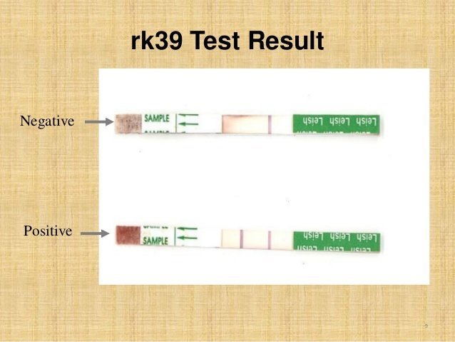
| Photo 2: rK39 Test kit result |
 |
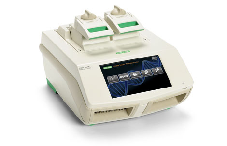
| Photo 3: Thermal Cycler |
 |
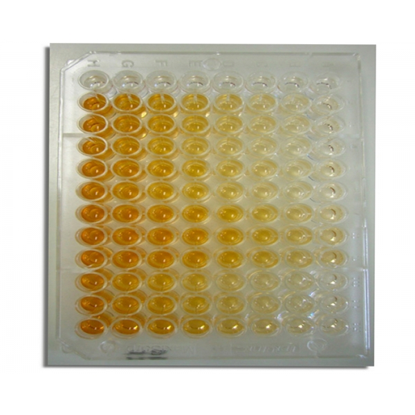
| Photo 4: ELISA Plate |
 |
| Photo 5: Fluorescence microscope |
 |
| Photo 6: Gene Xpert |
 |
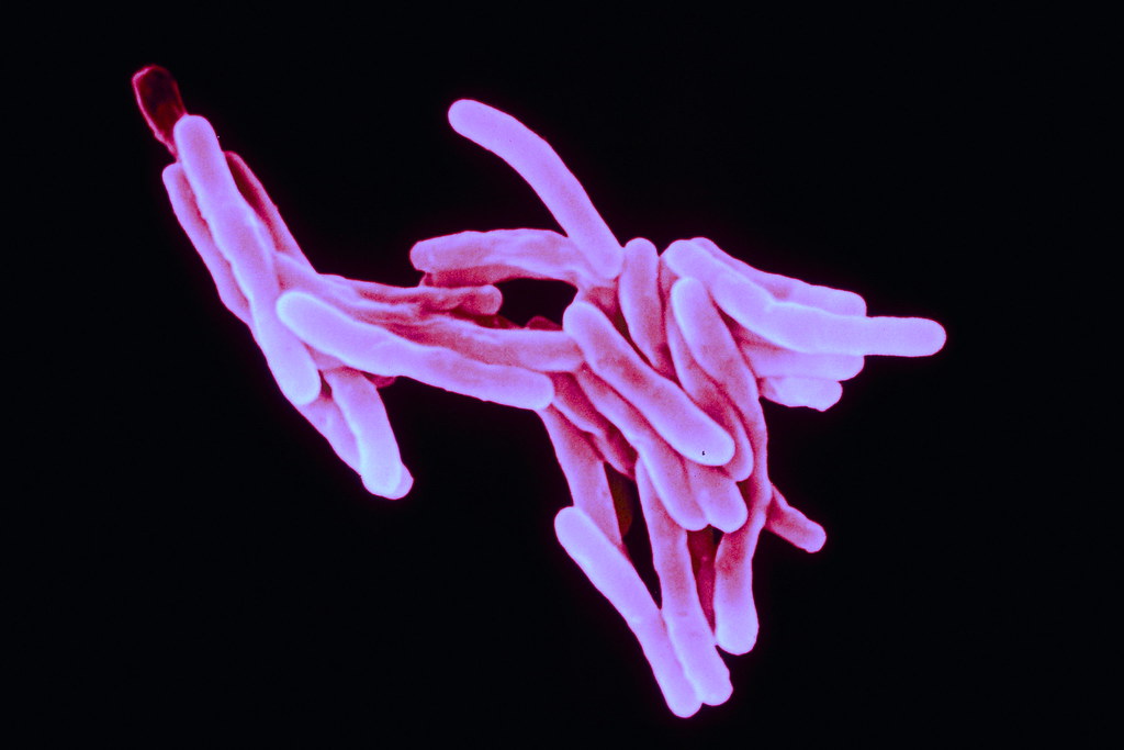
| Photo 7: M. tuberculosis |
Instrumentations
BPKIHS function as a center of excellence in the field of tropical and infectious diseases. Kala-azar Project in collaboration with international bodies and DOTS clinic in association/collaboration with the National Tuberculosis program and involving Municipality of Dharan and District Health Office, Sunsari with the application of following instrumentations.For Leishmaniasis
Laboratory diagnosis of leishmaniasis can be made by the following:
- demonstration of the parasite in tissues of relevance (spleen, liver, or lymph node tissue specimen) by light microscopic examination of the stained specimen, in vitro culture, or animal inoculation;
- detection of parasite DNA in tissue samples; or
- immunodiagnosis by detection of parasite antigen in tissue, blood, or urine samples, by detection of nonspecific or specific antileishmanial antibodies (immunoglobulin), or by assay for leishmania-specific
- cell-mediated immunity.
Specimens:
- tissue biopsy
- splenic aspirates
- bone marrow aspirates
Microscopy: Demonstration and isolation of parasite.
The commonly used method for diagnosing VL has been the demonstration of parasites in splenic or bone marrow aspirate. The presence of the parasite in lymph nodes, liver biopsy, or aspirate specimens or the buffy coat of peripheral blood can also be demonstrated. Amastigotes appear as round or oval bodies measuring 2 to 3 μm in length and are found intracellularly in monocytes and macrophages. In preparations stained with Giemsa or Leishman stain, the cytoplasm appears pale blue, with a relatively large nucleus that stains red. In the same plane as the nucleus, but at a right angle to it, it is a deep red or violet rod-like body called a kinetoplast.Laboratory Investigations and Diagnostic test:The following laboratory investigations and diagnostic tests will be done for diagnosis of Kala-azar and PKDL and be monitoring the progress and side effects of the drugs during the course of treatment.
Rapid dipstick test (rK39 test):
rK39 is a 39-amino acid repeat that is part of a kinesin-related protein in Leishmania chagasi and which is conserved within the Leishmania donovani complex. Among the available serological diagnosis of VL, only the immunochromatographic rK39 assay can be considered a point of care test for field application. They are currently the best available diagnostic tool for VL for use in remote areas, and their use in a field setting should be promoted. Apart from use in routine services, use of rK39 in campaigns and active case search is highly recommended. This test may be positive in a healthy person from the endemic areas (asymptomatic cases, past infection). Such a case should be followed up and treated only when they have indicative signs and symptoms of Kala-azar. rK39 is usually negative amongst PKDL with macular lesions and Leishmaniasis.
rK39 is available at Kala-azar endemic districts from level II and above health institutions.
Diagnosis of PKDL All suspected cases of PKDL should be confirmed with the rK39 test. However, it is important to note that the rK39 test may be negative in PKDL cases with only macular lesions and Kala-azar/HIV coinfection. If there is strong suspicion amongst rK39 negative suspected cases, such patients should be referred to centers where the diagnostic facility of PCR or demonstration of the parasite in pinch biopsy is available.
DNA detection method:
The development of PCR has provided a powerful approach to the application of molecular biology techniques to the diagnosis of leishmaniasis. Primers designed to amplify conserved sequences found in minicircles of KDNA of leishmanias of different species were tested in various tissues of relevance. Such a target was eminently suitable because the kinetoplast is known to possess thousands of copies of minicircle DNA.The PCR-ELISA technique can potentially be used for the diagnosis of VL from peripheral blood samples.
Contamination Control Program
- Space and time separation of pre and post-PCR activities
- Use of physical separation aids
- Use of ultraviolet (UV) light
- Use of aliquoted PCR reagents
- Incorporation of numerous positive and negative control blank
- Use of one or more various contamination control methods.
- Two broad methods of contamination control:
– Chemical methods that inactivate the amplicon’s ability to be templates in a new cycle of PCR.
In BPKIHS PCR lab use a spectrum of these methods to effectively control contamination.
Principle:
It is a chain reaction, a small fragment of the DNA section of interest needs to be identified which serves as the template for producing the primers that initiate the reaction. One DNA molecule is used to produce two copies, then four, then eight and so forth. This continuous doubling is accomplished by specific proteins known as polymerases, enzymes that are able to string together individual DNA building blocks to form long molecular strands. To do their job polymerases require a supply of DNA building blocks, i.e., the nucleotides consisting of the four bases adenine (A), thymine (T), cytosine (C) and guanine (G). They also need a small fragment of DNA, known as the primer, to which they attach the building blocks as well as a longer DNA molecule to serve as a template for constructing the new strand. If these three ingredients are supplied, the enzymes will construct exact copies of the templates.
Applications:
- Diagnosis of genetic diseases
- Genetic fingerprints
- Detection and diagnosis of infectious diseases
- Detection of infectious in the environment
- Personalized medicine
- PCR in research
- PCR is used in archaeology
Immunodiagnosis:
(i) Antigen detection.
Antigen detection is more specific than antibody-based immunodiagnostic tests. This method is also useful in the diagnosis of disease in cases where there is deficient antibody production (as in AIDS patients).A new latex agglutination test (KATEX) for detecting leishmanial antigen in urine of patients with VL. The antigen is detected quite early during the infection and the results of animal experiments suggest that the amount of detectable antigen tends to decline rapidly following chemotherapy. The test performed better than any of the serological tests when compared to microscopy.
(ii) Antibody detection.
The nonspecific methods, which depend upon raised globulin levels, have been used in the diagnosis of VL. Some of the tests used for detecting these nonspecific immunoglobulins are Napier’s formol gel or aldehyde test and the Chopra antimony test. Since these tests depend upon raised globulin levels, results can be positive in a host of conditions. Lack of specificity, as well as varying sensitivities, renders them highly unreliable.ELISA
ELISA has been used as a potential serodiagnostic tool for almost all infectious diseases, including leishmaniasis. The technique is highly sensitive, but its specificity depends upon the antigen used. Several antigens have been tried. The commonly used antigen is a crude soluble antigen (CSA).Specific antibodies can also be detected by Western blotting. For this type of testing, promastigotes of L. donovani are grown to log phase and lysed and the soluble protein is run on sodium dodecyl sulfate-polyacrylamide gel electrophoresis gels. The separated proteins were electroblotted onto a nitrocellulose membrane and probed with serum from the patient.
For Tuberculosis Diagnosis:
DOTS program is being run in association/collaboration with the National Tuberculosis program and involving Municipality of Dharan and District Health Office, Sunsari. The commitment of each stakeholder has been clearly spelled out. BPKIHS runs a DOTS clinic and provides the facility for the diagnosis, treatment, and management of complications. It also facilitates other DOTS sub-centers of Dharan Municipality for training and supervision. There are regular review meetings of the main center with the sub-centers, DHO and Dharan Municipality.Specimens:
The specimens for diagnosis of tuberculosis are,
- sputum
- urine
- CSF
- Stool
- Pleural fluid
- Lymph node biopsy
Microscopy:
The presence of acid-fast bacilli (AFB) on a sputum smear or other specimen often indicates TB disease. Acid-fast microscopy is easy and quick, but it does not confirm a diagnosis of TB because some acid-fast bacilli are not M. tuberculosis. Therefore, a culture is done on all initial samples to confirm the diagnosis. A positive culture for M. tuberculosis confirms the diagnosis of TB disease. Culture examinations should be completed on all specimens, regardless of AFB smear results.AFB staining:
When the smear is stained with carbol fuchsin, it solubilizes the lipoidal material present in the Mycobacterial cell wall but by the application of heat, carbol fuchsin further penetrates through the lipoidal wall and enters into the cytoplasm. Then after all the cell appears red. Then the smear is decolorized with the decolorizing agent (3% HCl in 95% alcohol) but the acid-fast cells are resistant due to the presence of a large amount of lipoidal material in their cell wall which prevents the penetration of decolorizing solution. The non-acid fast organism lacks the lipoidal material in their cell wall due to which they are easily decolorized, leaving the cells colorless. Then the smear is stained with counterstain, methylene blue. Only decolorized cells absorb the counterstain and take its color and appear blue while acid-fast cells retain the red color.Carbol fuchsin acid-fast stain or fluorescent auramine-rhodamine stain.
Fluorescent microscope:
The fluorescence microscope is an optical microscope that uses fluorescence to generate an image.Principle:
The fluorescence microscope is based on the principle of fluorescence. Fluorescence is a phenomenon in which certain fluorescent chemicals absorb the light of a particular wavelength and then emits the lights of larger wavelength with the lesser content of energy.
The specimen is illuminated with the light of a specific wavelength which is absorbed by the fluorophore, causing them to emit the light of longer wavelength. The excited light is separated from the emitted fluorescence through the use of spectral emission filter. The filters and diachronic mirror are selected to match with spectral excitation and the emitted fluorescence of the fluorophore used to label the specimen. In this way, the fluorescence of a single fluorophore is detected in the form of an image at a time. Multicolored images of several types of fluorophore can be detected by using a combination of the filter.
Application:
The fluorescence microscope is an important diagnostic tool for academic and pharmaceutical research, pathology, and clinical medicine.
Gene Xpert
The Genexpert test is a new molecular test for TB which diagnoses TB by detecting the presence of TB bacteria, as well as testing for resistance to the drug Rifampicin.The test is a molecular test which detects the DNA in TB bacteria. It uses a sputum sample and can give a result in less than 2 hours. it can also detect the genetic mutations associated with resistance to the drug Rifampicin.
Advantages
The main advantages of the test are, for diagnosis, reliability when compared to sputum microscopy and the speed of getting the result when compared with culture. For the diagnosis of TB, although sputum microscopy is both quick and cheap, it is often unreliable. It is particularly unreliable when people are HIV positive. Although culture gives a definitive diagnosis, to get the result usually takes weeks rather than the hours of the Genexpert test.
Summary and Conclusion:
BPKIHS Dharan runs along with the other hospital services and academic programs it is also the center of excellence in the field of tropical and infectious diseases. The ongoing Kala-azar Project in collaboration with international bodies and DOTS program in association/collaboration with the National Tuberculosis program and involving the Municipality of Dharan and District Health Office, Sunsari. For diagnosis and treatment of Kala-azar and Tuberculosis, various instrumentations such as PCR, Gene Xpert, Fluorescence microscope, Electrophoretic techniques are installed.We during our field visit observed the instrumentations, studied their principles and applications. Hence the field visit was found to be helpful and supportive to understand the various technique about different sample collection, instrumentation and their application for disease diagnosis and treatment.
References:
- www.bpkihs.edu
- www.mohp.gov.np
- www.pubmed.com
- Principles and techniques of Biochemistry and Molecular Biology
- MicrobiologyinPictures.com
Tags:
Microbiology

